#LIOS-004

SUMMARY
MitoCLue is a novel fluorescent probe that enables 3 complementary cardiolipin (CL) assays – binding affinity measurement, CL quantitative analysis, and CL imaging. Designed for both mitochondrial research and drug safety profiling, MitoCLue supports drug discovery with high-throughput, reproducible and biologically relevant results.
BACKGROUND
Cardiolipin (CL) is a key phospholipid responsible for mitochondrial function and structural integrity. Its depletion is a hallmark of many diseases such as heart failure, diabetes, and aging. While CL is a promising target for mitochondria-specific drugs and drug delivery systems, off-target interactions can cause drugs to accumulate in mitochondria, potentially leading to serious side effects. Until recently, there was no unified assay that would allow to estimate compounds` affinity to CL in a high throughput screening.
FEATURES AND KEY BENEFITS
1. Binding Affinity Assay
Features
- Validated in 96-well format (Z’ > 0.7), adaptable to 384-well
- EC₅₀ determination across 6–7 log concentration range
- Suitable for artificial membranes models or mitoplasts
Benefits
- Screening of compound libraries for CL binding
- Facilitates early-stage drug discovery and safety profiling
- Standard plate reader compatible
2. CL quantitative analysis
Features
- Direct quantification of CL in isolated mitochondria
Benefits
- Comparative analysis of CL levels across tissues or conditions
3. CL imaging
Features
- CL-specific probe suitable for confocal microscopy
Benefits
- Visualizes mitochondrial distribution and CL-rich zones
- Non-toxic to cells
REFERENCES
[1] P. Dimitrijevs, P. Arsenyan, Sensors&Actuators B: Chem., 346, 2021, 130537; doi: 10.1016/j.snb.2021.130537 .
[2] P. Dimitrijevs, I. Domracheva, P. Arsenyan, New J. Chem., 2020, 44(23), pp. 9626–9633; doi: 10.1039/D0NJ02116D.
[3] P. Dimitrijevs, et al., Lipids Health Dis. 2025, 24, 76. doi: 10.1186/s12944-025-02499-5.
[4] C. Pegoraro, et al., Adv. Mater. 2025, 37, 2411595; doi: 10.1002/adma.202411595.
AVAILABLE
for licensing, service or research collaboration.
CONTACT
Dr. Anna Stikāne: anna.stikane@osi.lv
Latvian Institute of Organic Synthesis, Aizkraukles Str. 21, Riga LV-1006, Latvia
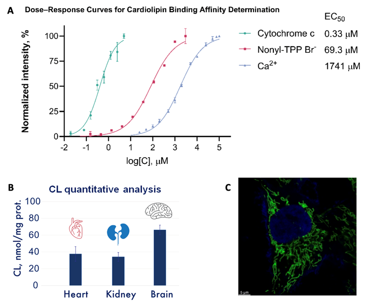
A. Dose–response curves of cytochrome c, nonyl(triphenyl)phosphonium bromide, and Ca²⁺ demonstrating their relative binding affinities to CL as measured by the MitoCLue assay.
B. Total CL content in mitochondrial fractions isolated from mouse heart, kidney and brain.
C. Confocal microscopy images of a live A2058 cell stained with MitoCLue (green) and Hoechst (Blue).